Toshiba's VISIONS Magazine - issue 26
•
3 likes•1,576 views

VISIONS magazine is a publication of Toshiba Medical Systems Europe (Toshiba), and is offered free of charge to medical and health professionals. The magazine is published twice a year but is also available as an online portal. News items and articles are announced, pre-publication, via social media in dedicated groups. Toshiba stores data of readers and users, as far as known, of the online VISIONS portal. This data is used to send out the magazine and inform about new developments in the clinical market. Users can customize their preferences or opt-out in the online VISIONS profile at www.toshiba-medical.eu/visions. ©1999-2016 by Toshiba Medical Systems Europe. All rights reserved

Recommended
More Related Content
What's hot
What's hot (8)
Viewers also liked
Viewers also liked (14)
Similar to Toshiba's VISIONS Magazine - issue 26
Similar to Toshiba's VISIONS Magazine - issue 26 (20)
More from Canon Medical Systems Europe
More from Canon Medical Systems Europe (16)
Recently uploaded
Recently uploaded (20)
Toshiba's VISIONS Magazine - issue 26
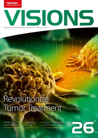
- 1. SEMAR in Neuroradiology Rivierenland Hospital Lung SURESubtraction in Everyday Practice Revolutionize Tumor Treatment VISIONSMagazine for Medical & Health Professionals I February 2016 24 I COMPUTED TOMOGRAPHY 10 I MODALITY FUSION 33 I CUSTOMER FOCUS 55 I COMPUTED TOMOGRAPHY 26
- 2. VISIONS magazine is a publication of Toshiba Medical Systems Europe (Toshiba) and is offered free of charge to medical and health professionals. The magazine is published twice a year. Registration to access full, previously published, digital editions can be done via the web site: www.toshiba-medical.eu/visions. Toshiba stores and uses personal data of the registration to send out the magazine and inform about new developments in the clinical market. Readers can customize preferences or opt-out, after registration, in the online VISIONS profile. News items and articles are announced firstly, as pre-publication, via the dedicated VISIONS LinkedIn Group: https://www.linkedin.com/groups/3698045. In this group you can also actively participate in discussions about the content and future direction of the magazine. Alternatively you can e-mail us at: VISIONS@tmse.nl. Follow us also on SlideShare: http://www.slideshare.net/toshibamedical. Publisher TOSHIBA Medical Systems Europe B.V. Zilverstraat 1 NL-2718 RP Zoetermeer Tel.: +31 79 368 92 22 Fax: +31 79 368 94 44 Web: www.toshiba-medical.eu Email: info@tmse.nl Editor-in-chief Jack Hoogendoorn (jack.hoogendoorn@toshiba-medical.eu) Modality coordinators and reviewers CT: Roy Irwan, Chloe Steveson UL: Jeroen Uijttenhout Design & Layout Boerma Reclame (www.boermareclame.com) Commissioned photography Cojan van Toor (www.cojanvantoor.nl) Printmanagement Het Staat Gedrukt (www.hetstaatgedrukt.nl) Text contributions “Customer Focus: Rivierenland Hospital, Tiel” by The Creative Practice (www.thecreativepractice.com) Subscription Service www.toshiba-medical.eu/visions VISIONS magazine is covering Toshiba’s European region and as such reflects products, technologies and services for this particular area. The mentioned products may not be available in other geographic regions. Please consult your Toshiba representative sales office in case of any questions. © 2016 by TOSHIBA Medical Systems Europe All rights reserved ISSN 1617-2876 26 Partnering advanced diagnostic imaging with minimally invasive intervention is possibly the most promising future for radiology and surgery in oncology. Read more on page 10. Digital illustration of Cancer cell in colour background”, ID 45675334 © Krishnacreations | Dreamstime.com www.toshiba-medical.eu ULTRASOUND CT MRI X-RAY SERVICES Our lives and social environment are subject to constant change and create ever-increasing needs and high demand for better medical solutions. We at Toshiba aim to maximize the quality, safety, and efficiency of medical care, supporting clinical practice with reliable quality products and innovative, cutting-edge technologies. The high image resolution and superior operability of our medical systems creates new clinical value. While our advanced applications, supported by highly reliable technologies, open the door to the next stage of medical care. We will continue to provide a wide variety of leading-edge solutions for the benefit of all people around the world, and seek to further development in the field of healthcare following our basic commitments: “Improving the quality of life”, “Lifelong commitment to innovation”, and “Achieving lifetime partnerships”. Toshiba: Made for Life! MADE FOR LIFE.
- 3. VISIONS26 | 3 We live in an era that is characterized by rapid change. Change in itself is not new, but perpetual, as Heraclitus (535-475 BC) already observed: Everything changes and nothing remains still ...You cannot step twice into the same stream1 However, the pace of change these days is undeniably different. Computing power has doubled every two years2, while products are becoming smaller and smarter. Our vision and interests are no longer limited to our immediate region, but extend over the entire planet by leveraging communication tools, such as satellite communications, the Internet and social media. While “one small step for man” on the moon has long been relegated to history, the next “giant leap for mankind” focuses on human settlement on Mars, and self-propelled vehicles are almost a reality. Such acceleration is also seen in high-tech industries, in which the technologies used in today’s products, renew, improve and merge continuously at a rapid pace. In the medical imaging industry, advanced technologies enable faster, safer and more extensive completion of daily routines and complex work flows. Here, modality fusion can provide an ever better opportunity for more accurate diagnoses. Toshiba’s ‘intellectual brains’work ‘twenty-four-seven’ on futuristic solutions to progress developments and ensure that what was not visible or available yesterday, appears as a reality on our horizon today. Obviously, organizational management is also subject to change in these dynamic conditions. Partnerships, mergers and acquisitions are realistic options, whereas continuity and other guarantees for the future are unmistakable key focus areas. What exact changes the future holds for us are unknown. What I do know is that our unprecedented commitment, personal attention, customer-centered mentality, specialized skills and knowledge will remain. These specific qualities are deeply anchored in our corporate culture and employees, and are characteristic and distinctive of Toshiba as your Dedicated Imaging Specialists. Kind regards, Dear reader, EDITORIAL Jack Hoogendoorn Sr. Manager Marketing Communications Toshiba Medical Systems Europe BV 1 Quoted by Plato in Cratylus, 402a 2 Moore’s Law
- 4. ©2016 TOSHIBA MEDICAL SYSTEMS4 | VISIONS26 10 Revolutionize Tumor Treatment 18 New frontiers in forensic radiology 22 Bismuth Shields: Are they necessary in 2016? 24 SEMAR in neuroradiology 28 Seeing the unseen 33 Rivierenland Hospital, Tiel 43 Providing New Diagnostic Possibilities in Tanzania 10 28 33 Fused advanced diagnostic imaging combined with minimally invasive intervention. 3D SMI enables visualization of the entire vascular structure in an area of interest. The Radiology Department at Rivierenland Hospital, Tiel, The Netherlands CONTENTS COMPUTED TOMOGRAPHY CUSTOMER FOCUS ULTRASOUND COMPUTED TOMOGRAPHY ULTRASOUND COMPUTED TOMOGRAPHY MODALITY FUSION CAD, CTA, Adaptive Motion Correction (AMC), Forensics Tumor treatment, cancer, tumor ablation Bismuth shields, image quality Single energy, Metal artefact reduction SMI, Doppler Corporate Social Responsibility, Tanzania VISIONS SPECIAL 1 cm
- 5. VISIONS26 | 5 46 58 51 51 The Aquilion Lightning sets the standard in the 16-slice class of CT systems with excellent specifications and modern clinical applications. Dual Energy technology provides differentiation of tissues of similar density and atomic number. Noninvasive quantification of left ventricular (LV) contractility is a topical challenge in modern echocardiography. Noninvasive quantification of left ventricular (LV) contractility is a topical challenge in modern echocardiography. COMPUTED TOMOGRAPHY 46 Aquilion Lightning: world’s first 16-row low-dose routine scanner 51 Normal values of left ventricle strain using 3D Wall Motion Tracking technology 55 Lung SURESubstraction in Everyday Practice 58 Practical uses of Dual Energy CT 62 Determination of the cerebral perfusion territories using CT Perfusion imaging 03 Editorial 06 News 09 Message from the President ULTRASOUND Left ventricle, strain ratio, 3D wall motion tracking Routine Radiology, dose reduction, AIDR 3D Enhanced, 78 cm gantry opening COMPUTED TOMOGRAPHY COMPUTED TOMOGRAPHY COMPUTED TOMOGRAPHY Dual energy CT Perfusion, perfusion territories, CAD CTPA, Lungs, Subtraction
- 6. ©2016 TOSHIBA MEDICAL SYSTEMS6 | VISIONS26 NEWS Olea Medical to join Toshiba Medical Systems Corporation Group New edition of the Vantage Titan 3T FIRST enhances images quality and lowers dose Wearvue: a small, light, next genera- tion wearable work support tool The acquisition enables Toshiba to accelerate the growth of its MRI business and offer new clinical added value to healthcare providers by leveraging Olea’s cutting-edge software technology for advanced post-processing and image analysis as well as its broad relationships with the world’s key research institutions and customers. Toshiba will also be able to provide multi-modality solutions by integrating Olea’s post-processing software with “Vitrea”, a widely known enterprise and scanner-connected workstation by Vital Images Inc., another subsidiary of Toshiba, to improve collaboration among experts, consolidate and standardize workflows and enhance diagnostic confidence. More information: http://www.olea-medical.com/en Toshiba has launched the Toshiba 3-tesla MRI system Vantage Titan™ 3T / iS Edition (iS: intelligent Solution). In order to fully satisfy the clinical demands, the system incorporates the new gradient coil system employed in Saturn Gradient, which enables higher image quality with open-bore MRI systems. It also provides a suite of advanced applications, achieving robust, efficient, and highly accurate examinations in the safe and reliable environments. More information: http://tinyurl.com/o957wvtClinically viable model-based iterative reconstruction is now a reality with the launch of Toshiba’s FIRST; Forward projected model-based Iterative Reconstruction SoluTion. FIRST improves image quality with significant noise reduction while reducing radiation dose and drastically cuts the time needed for model- based CT image reconstruction, helping providers make critical diagnoses and treatment decisions more effectively. AvailablefortheAquilionONE™FamilyofCTsystems,FIRSTimproves high-contrast spatial resolution and helps to make exams safer for patients by providing reduction in dose, meaning customers don’t need to choose between safety and high quality images. More information: http://tinyurl.com/hwceylk “Wearvue”is a next generation wearable device for the B2B market that will help to achieve a hands-free working environments in facilities such as factories and logistics centres as well as during activities like infrastructure maintenance. Wearvue is worn like a pair of glasses and, at only 50 grams, is designed to be light and comfortable enough for long periods of use. The design also achieves a natural appearance that does not distract nor disturb people during conversations. In use, aToshiba- developed original optical system projects data, text and colour images, to the right-hand lens of Wearvue, with a 1:1 aspect ratio. The optical system secures a wide viewing area and a depth of field that makes the data easy to see without obscuring the wider view. Wearvue also includes a personal adjuster for changing the angle of image projection, ensuring that it can be worn by an estimated 98% of Japanese adults.
- 7. VISIONS26 | 7 NEWS AMSTERDAM AMSTERDAM AMSTERDAM A NTTDoCoMoYoyogiBuilding,Tokyo A Landmarktower,Yokohama B Asakusatemple,Tokyo C Tokyotower,Tokyo D ToshibaHeadquarter,Tokyo E Odaiba'sstatueofliberty,Tokyo F FujiSankeiBuilding,Tokyo G Cosmoclock21,Yokohama H Ginza'swako,Tokyo I Tokyoskytree,Tokyo J ToshibaMedicalSystemsHeadquarter,Tokyo K ShinjukuCenterBuilding,Tokyo L B C D E F G H I J K 1 2 3 4 5 6 Koningshavenbrug,Rotterdam OudeKerk,Amsterdam Vredespaleis,DenHaag BasiliekvandeH.Nicolaas,Amsterdam DeMunt,Amsterdam Hoftoren,DenHaag Binnenhof,DenHaag 1 2 3 4 5 6 7 7 8 9 10 11 13 Euromast,Rotterdam HetPaleisopdeDam,Amsterdam Erasmusbrug,Rotterdam NEMO,Amsterdam Domtoren,Utrecht HotelNewYork,Rotterdam ToshibaMedicalSystems,Zoetermeer 8 9 10 11 12 13 14 14 12 ► Next page is part of the VISIONS Photo Page Series reflecting an eye for the beauty of our planet, the environment and the direct surroundings where Toshiba’s systems are installed by Toshiba and its customers. Not the actual imaging products but photos of sceneries, cities, countries or other cultural aspects are highlighted on this photo page. The Photo Page is based upon an idea of Prof. Edwin van Beek. Every reader of VISIONS can participate and get their picture published. The submitted content should include: high resolution (300dpi) image, photo of the hospital and a brief text, name of photographer and Toshiba system(s) installed. The complete result is shown on the opposite page. Send your pictures and texts to: jack.hoogendoorn@toshiba-medical.eu, Subject: Photo Page Meet us in Tokyo or Amsterdam It is with great pride thatToshiba announces the new high tech training facility; theTokyo room.The design of the Tokyo room is based upon Toshiba’s new view on learning development.The many technological features and unique lay-out adds a new dimension to the training courses, making them even more valuable than so far. The room is easy recognizable by a stylish and detailed silhouette of the city’s skyline. The next room where the skyline-concept will be applied is the Amsterdam room. Actually a large area where the usage of state-of-the-art X-ray systems are taught and demonstrated. Both skylines include some recognizable and well known highlights. How good is your ‘skyline- knowledge’? Do the test!
- 8. ©2016 TOSHIBA MEDICAL SYSTEMS8 | VISIONS26 Ego Vivo/Self-portrait 25 was shown at Lowlands; an annual three day music and performing arts festival, held in the Netherlands in August since 1993. Although the main focus is on music - rock, pop, dance, hip hop and alternative - Lowlands also offers cinema, (street) theatre, cabaret, art, science, stand-up comedy, ballet and literature. Lowlands is attended by around 50,000 visitors, spread over 250 acts and more than ten stages every year. Text Source: Wikipedia – Photography: Annick Vroom Ego Vivo/Self-portrait 25, 2013 Bronze and Concrete by artist Caspar Berger, is part of the ongoing Skeleton project, started in 2012 Caspar Berger: “In the Skeleton project I want to make tangible what lies beneath the skin. I have had my entire body scanned using the Toshiba Aquilion Prime 80-slice CT-scanner to produce, with a 3D printer, the most accurate possible copy of my skeleton. I can then make silicone moulds allowing me to cast copies of my skeleton in bronze, silver and plaster.” Text Source: Caspar Berger (www.casparberger.nl) Photography: Bas Uterwijk (www.basuterwijk.com)
- 9. VISIONS26 | 9 PRESIDENT’S MESSAGE “We are, and always will be, committed to you and your business” You may have heard recently that Toshiba Corporation is restructuring and seeking a majority shareholder(s) for Toshiba Medical Systems Corporation, headquartered in Japan. I would like to use this opportunity to reaffirm our commitment to you and the continuation of our services and support you have come to rely upon. Toshiba is undertaking this step to strengthen the financial resources dedicated towards realising our company’s medical growth strategies. It is a decision based on careful and thorough consideration. We have been contributing to the medical industry by providing innovative and advanced products, solutions and services to our customers world-wide for over 100 years. However, today’s industry is growing and changing rapidly. Despite the fact that Toshiba Corporation has invested significantly over the years in healthcare, in order us to reach our full future potential, continued investment is required, particularly in the areas such as research and development and mergers and acquisitions. Inviting outside investments in the business by majority shareholder(s) will allow us to make timely strategic investments that would lead to higher growth. We see Toshiba Corporation’s recent decision as a positive one for our future to continue setting high value on customer satisfaction and providing advanced products and solutions including service and maintenance support in ways that demonstrate our commitment. We are expecting to continue our current activities and direction focused on strong growth in de medical imaging market. The sales, marketing, installation, training, maintenance and service of our products will continue. Over the mid/long-term we are looking forward to expanding our business by strengthening RD, introducing leading-edge technology and adding new business segments to our portfolio. I assure you that our commitment and dedication to you and your business will stay unchanged. I would like to thank you for your patience and understanding during this time of transition. Toshio Takiguchi President and Chief Executive Officer Toshiba Medical Systems Corporation
- 10. To put it briefly, fusion technique in radiology means simultaneously using images taken with different modalities (ultrasound, CT, MRI, PET-CT) and at different times for the same patient. The images can be matched, and/or in some cases fused. Our team at Danderyd Hospital uses two types of fusion techniques to get closer to a desirable and optimal result. One is fusion of ultrasound with CT or MRI, and the other is fusion of CT with CT, in 2D and 3D. Toshiba’s ultrasound system (Aplio 500) and Aquilion ONE CT scanner are used at Danderyd Hospital both for diagnostic and intervention purposes. We work intensely with the development of tumor diagnostics before and after ablation treatment, and with developing more precise placement of needles/electrodes for thermal (MW, RF) and non thermal (IRE) ablation treatment. We are also working on improving the work flow surrounding ablation treatments. The results are very positive and we presented our experiences at a symposium during a scientific conference held atThe Swedish Society of Medicine in Stockholm.” What is the most promising future possibility for radiology and surgery in oncology? The answer is clear: Partnering advanced diagnostic imaging with minimally invasive intervention. That is, if you ask radiologist Anastasios Michos, Deputy Department Head and Method Development Specialist for ablation therapy within the Radiology Department of Danderyd Hospital, Stockholm, Sweden. Anastasios Michos 1) TECHNOLOGY MODALITY FUSION 1) Deputy Department Head and Method Development Specialist for ablation therapy within the Radiology Department of Danderyd Hospital, Stockholm, Sweden. ”The two fields are directly connected. If advanced diagnostic imaging and the effort to achieve minimally invasive intervention are developed in harmony with the needs of modern healthcare, a real revolution in tumor treatment will be possible.” Another important parameter is the cooperation between healthcare representatives, such as hospital administration, physicians and nurses, and the medical industry. These parties could - and should - collaborate, with the joint goal of helping cancer patients and opening up new paths in cancer diagnostics and treatment. At Danderyd Hospital we started a project a few years ago specifically on the theme “advanced diagnostic imaging and minimally invasive intervention”, where the main goal is to develop the techniques for tumor ablation. Our team, the so called ablation team, consists of radiologists, surgeons and anesthesiologists as well as industry representatives, mainly from Toshiba, Vital Images and Angiodynamics. Tumor treatment, cancer, tumor ablation Revolutionize Tumor Treatment Combining Advanced Diagnostic Imaging and Minimal Invasive Intervention ©2016 TOSHIBA MEDICAL SYSTEMS10 | VISIONS26
- 11. Following are some cases where ultrasound fusion was used to perform diagnostic and/or interventional examinations. GNEU150013 Kidney Tumor Diagnostics and Kidney Tumor Ablation Guided by Ultrasound-CT Fusion Small exophytic kidney tumor. The tumor is localized with the help of ultrasound-CT fusion technique and thereafter ultrasound contrast. Image 1 shows the abnormality loaded with ultrasound contrast medium. Image 2 shows a corrected match between CT and ultrasound images. Image 3: The radiofrequency needle is placed with support from the virtual needle guide navigation and the tumor is ablated. Image 4: Vitrea software (ViTAL Images) is used real-time during the procedure for Fusion and 3D reconstruction of CT with the needle in place. With the software the tumor (yellow), kidney (blue) and needle are visible, isolated Image 3 Image 1 Mr. Anastasios Michos demonstrating fusion technology on a phantom model The screen of the Aplio 500 shows the needle position in both the Ultrasound and CT image Image 2 Smart Fusion: Correlating different imaging modalities in real-time, Smart Fusion allows you to locate difficult lesions faster, to navigate complex anatomy securely, and to improve your confidence while carrying out invasive procedures. For a comprehensive pre- and post-interventional evaluation Smart Fusion allows you to work in any ultrasound imaging mode including color Doppler and contrast-enhanced ultrasound. Smart Fusion reads 3D DICOM data sets from all major imaging modalities and shows the corresponding images contained in real-time adjacent to the live ultrasound display. VISIONS26 | 11
- 12. Image 6 Image 4 Image 5 Biopsy Guided by Ultrasound-CT Fusion Lung Biopsy With the help of ultrasound-CT fusion a small peripheral change of the lung tissue is localized. After contrast- enhanced ultrasound to identify any necrotic areas a safe lung biopsy can be performed. from the CT image. Furthermore all images are fused and a 3D reconstruction is created showing clearly that the needle is in the desired position relative to the tumor. Image 5 shows a CT image of the tumor before treatment and image 6 shows a CT image of the tumor three months after treatment (an ablation cavity has appeared that covers the entire tumor). ©2016 TOSHIBA MEDICAL SYSTEMS12 | VISIONS26
- 13. Small suspected liver change in a patient with colon cancer. The change is visible on CT but not on ultrasound. With the help of ultrasound-CT fusion followed by contrast ultrasound the change can be localized anatomically (see green circle, image 1). The change is isoechogenic in the arterial phase (image 2) and has wash-out in the portal phase (image 3); therefore a metastasis is strongly suspected. Small tumorous change in kidney (see green circle on MRI image) to be ablated. The change is not visible on ultrasound. With the help of ultrasound-MRI fusion the change can be localized anatomically (image 1). Through contrast-enhanced ultrasound it can be visualized (green circle, image 3). The radiofrequency needle can then be placed and ablation can be performed. Tumor Diagnostics With the Help of Ultrasound-CT/MRI Fusion Liver Tumor Diagnostics Kidney Tumor Diagnostics Image 2 Image 3Image 3 Image 2 Image 1 Image 1 GNEU150013 VISIONS26 | 13
- 14. Irreversible Electroporation (IRE treatment - Nanoknife) of Liver Tumor Atumor,justunderthreecentimeterswide,closetothecranial part of the gallbladder and in direct contact with the portal vein (see image 1). To avoid risking damage to blood vessels and/or gallbladder, the decision is made to use IRE ablation technique. Seven IRE needles (six around the tumor and one centrally) are positioned with the help of Toshiba’s Aplio 500 ultrasound system; the distance between the needles should be somewhere between one and two centimeters. After needle placement the position is verified using the Aquilion ONE CT scanner (AIDR 3D)without contrast (see image 2). Fusion and 3D reconstruction of CT with contrast before needle placement and CT without contrast with the needles in place (see images 3-6). Since CT verification after needle placement (image 2) is done without contrast it cannot give Image 2 Image 4 Image 6 Image 1 Image 3 Image 5 sufficient information regarding needle position in relation to the tumor and blood vessels. To solve this problem Vitrea software (Vital Images) is used live during the procedure. With the help of this software: - tumor and liver are isolated from image 1 - needles are isolated from image 2 - all images are fused and a 3D reconstruction is created. These 3D fused images show that the needles are in a good position relative to the tumor and blood vessels, albeit somewhat deep. All needles are backed up a bit and the treatment is performed.The treatment consists of 90 electric pulses of 3000 volts between each needle pair, and takes around half an hour in total. ©2016 TOSHIBA MEDICAL SYSTEMS14 | VISIONS26
- 15. GNEU150013 Image 1 shows an ultrasound image of the tumor without contrast before treatment and image 2 shows it after treatment. Image 3 shows an ultrasound image of the tumor with contrast after treatment. Image 1 shows a CT image of the tumor before treatment (here it can be seen that the tumor is on the portal vein and is partially compressing it). Image 2 shows a CT image of the tumor two weeks after IRE treatment (in the tumor location Image 1 Image 2 Image 3 Image 3Image 2Image 1 an ablation cavity has appeared that covers the entire tumor; the portal vein is open). Image 3 shows a CT image of the tumor three months after treatment (the ablation cavity has shrunk; the portal vein is open). VISIONS26 | 15
- 16. Small liver metastasis which is poorly visualized with ultrasound. The metastasis can be localized anatomically with the help of ultrasound-MRI fusion (see yellow circle). The microwave needle must be placed carefully since the abnormality is situated deep in the tissue and there are a few blood vessels along the way that must be avoided. The virtual needle guide navigation technique is used to obtain better control during placement (the green line and the little blue dot show location of the needle and the needle point respectively, the purple oval shows the estimated result of the ablation). Liver metastasis which is poorly visualized with ultrasound. The metastasis can be localized anatomically with the help of ultrasound-MRI fusion (see yellow circle). The microwave needle must be placed carefully since the abnormality is situated deep in the tissue (close to the cardia, see pink circle) and there are a few blood vessels along the way that must be avoided. The virtual needle guide navigation technique is used to obtain better control during placement (the green line and the little blue dot show location of the needle and needle point respectively). The purple oval shows the estimated result of the ablation. Image 2 shows ongoing ablation after correct needle placement. Liver Tumor Ablation Guided by Ultrasound-CT/MRI Fusion and Virtual Needle Guide Navigator Case 1 Case 2 Image 1 Image 2 ©2016 TOSHIBA MEDICAL SYSTEMS16 | VISIONS26
- 17. Get even smarter! Register now for this unique 2-day workshop on Neurological Imaging with Dynamic Volume CT. The workshop aims to provide a working knowledge of current 320-row dynamic volume MDCT (Aquilion ONETM ViSION Edition) for neurological imaging, covering evaluation of both brain perfusion and dynamic CTA, as well as cerebrovascular anatomy and pathology. An experienced faculty will guide you from scan procedures to the implementation of comprehensive image protocols for diagnosis and management of neurological conditions. In addition, the highly interactive program allows hands-on interpretation and discussion of clinical case studies.Afterwards, participants will know how to apply dynamic volume CT for optimal brain imaging. 09:00 - 09:10 Opening and introduction 09:10 - 09:50 Imaging of ischemic stroke: what is important and why? Marianne van Walderveen 09:50 - 10:15 Basic principles of perfusion Uulke van der Heide 10:15 - 10:45 Coffee 10:45 - 11:15 Brain perfusion scan procedures and analyses Joost Roelofs 11:15 - 12:00 Comprehensive ischemic stroke protocol: tips and tricks for the clinical practice Marianne van Walderveen 12:00 - 12:30 CT technology and radiation dose Raoul Joemai 12:30 - 14:00 Lunch 14:00 - 14:15 Workstation introduction Joost Roelofs 14:15 - 17:00 Hands-on workstation with clinical cases, read with the experts 19:00 - 22:00 Dinner 09:00 - 09:45 Imaging of fistulous intracranial lesions, what is important and why Patrick Brouwer 09:45 - 10:05 4D CTA acquisition protocol, how and why? Joost Roelofs 10:05 - 10:30 4D CTA, technical possibilities, pitfalls and artifacts Peter Willems 10:30 - 11:00 Coffee 11:00 - 11:20 4D CTA in AVMs Peter Willems 11:20 - 11:40 4D CTA in dAVF Peter Willems 11:40 - 12:00 4D CTA in miscellaneous clinical conditions and future applications Patrick Brouwer 12:00 - 13:30 Lunch 13:30 - 15:45 Hands-on workstation with clinical cases, read with the experts 15:45 - 16:00 Course Diploma and Adjourn First Day Brain Perfusion/Dynamic CTA Second Day 4D CTA We look forward to welcoming you to Leiden! Please register at: http://bit.ly/1ZlWNXj Neurological Imaging with Dynamic Volume CT 12 - 13 May 2016
- 18. ©2016 TOSHIBA MEDICAL SYSTEMS18 | VISIONS26 The increased knowledge and improved technology in Radiology facilitated the use of diagnostic imaging in forensic settings. In 1896, thanks to the brilliant idea of a coroner, in court a radiographic film was used to document the anatomical location of a bullet wound following a gunshot. Later, in the wake of this unusual application of diagnostic imag- ing, the use of radiology in the field of forensics gained enthusiastic endorsements. In 1984 in Italy, Di Maio published a handbook on the correct use of diagnostic imaging in the study of deaths by firearms, in order to assist and to guide traditional autopsy. A traditional autopsy provides extensive information for the patholo- gist, however a radiological ‘pre-autopsy’ may be able to guide and direct the traditional autopsy. In 1983 the first post-mortem computed tomography (PMCT) was made. A PMCT satisfies three fundamental objectives of a post- mortem examination: the identification of the corpse, finding any foreign bodies and assessment of the cause of death1. In clinical practice the physician determines the diagno- sis and choice of treatment strategy based on both the radiological report and a physical medical examination since Radiology can never completely replace the physi- cal examination. The same applies to forensics, where a non-invasive “pre-autopsy” guides coroner in the search of the main elements that could cause death, but does not replace the actual physical examination, the autopsy TECHNOLOGY CAD, CTA, Adaptive Motion Correction (AMC), Forensics New frontiers in forensic radiology Since ancient times people have tried to find answers to the question that always accompanies man’s life: the way of death. Until recently traditional autopsy was the only method to identify the cause of death. However, the sense of respect for the corpse and the fear of desecration represented serious obstacles to the spread of this practice. COMPUTED TOMOGRAPHY 1) Department of Radiology, University of Foggia, Viale Luigi Pinto 1, 71100 Foggia, Italy. 2) Department of Forensic Pathology, University of Foggia, Colonnello d’Avanzo Hospital, Viale degli Aviatori 1, 71100 Foggia, Italy. 3) Department of Radiology, Scientific Institute Hospital “Casa Sollievo della Sofferenza”, Viale Cappuccini 1, 71013 San Giovanni Rotondo, Foggia, Italy. Gianpaolo Grilli 1) Giulio Zizzo 1), Annagrazia Cecere 1), Cristoforo Pomara 2), Giuseppe Guglielmi 1,3) Dr. Gianpaolo Grilli Dr. Giulio Zizzo Dr. Giuseppe Guglielmi in this case. This innovative technique was commonly called “Virtopsy” because it combines two fundamental aspects in the study of the corpse. In fact, “virtus” reflects the need to make a useful and efficient investigation and “autopsy” refers to describe something that you see with your own eyes. The combination of the two words sug- gests the radiologist will substitute the subjectivity associ- ated with the activity of“seeing with their own eyes”with the execution of an exam that documents impartially the anatomical lesions responsible of the death. Compared with conventional autopsy, Virtopsy with CT has many advantages. First, the traditional autopsy is interpreted in many cultures (Judaism Islam) as an attempt to desecrate the corpse and, for this reason, it is rejected. Additionally, a PMCT is non-invasive, so doesn’t interfere with the corpse allowing a traditional autopsy to be performed afterwards with only minimal disruption. The possibility to give a high dose of radiation permits a large amount of information to be acquired for further detailed analysis at a later date after the corpse has been buried. In contrast, the traditional autopsy doesn’t allow further study into the cause of death at a later time because the collection of biological materials can only occur during the autopsy. PMCT also allows evaluation of the presence of bodily gas and foreign bodies which are not possible with a traditional autopsy. Despite all these indisputable advantages, Virtopsy doesn’t allow a “color” assessment of anatomic lesions: such a drawback could be solved with the application of 3D volume rendering (3D VR) techniques. Furthermore, it doesn’t allow the differentiation of soft tissues, which is better assessed with magnetic resonance imaging (MRI), and evaluation of vascular lesions, that would require the administration of a contrast agent. In view of these con- siderations, PMCT has most use in cases of identification of the corpse, multiple trauma, drowning, burns, gunshot injuries, hanging and documentation of previous injuries. Deaths from cardiac causes deserve special treatment, because the main cause of natural death is heart failure.
- 19. Figure 1a: Post mortem CT (PMCT) shows soft tissue swelling of right neck Figure 1b-c: PMCT-angiography (PMCTA) showing the leakage of contrast agent into the trachea (2b) and the peri-pharyngeal spaces (2c) in multi planar reformats (MPR) VISIONS26 | 19CTEU1500105 For this reason, it’s very important to differentiate the cardiac lesions responsible for death from concomitant injuries which are not sufficient in themselves to cause death. The further development of PMCT techniques led to the introduction of contrast agents to assess vascular lesions and post mortem CT angiography (PMCTA)2. The use of contrast agents, permits the complete filling of the vascular system and helps to identify vascular lesions and other sources of bleeding causing death. The standard protocol consists in a non contrast acqui- sition followed by three acquisitions with contrast. An imaging finding is considered real when it is present in at least two of the three post-contrast phases; other- wise it is interpreted as an artifact. The contrast agent, consisting of paraffin oil and 6% of Angiofil® (Fumedica AG, Muri, Switzerland), it is usually injected into the femoral vessels through the heart-lung machine, Virtangio® perfusion device (Fumedica AG). The com- parison between the traditional autopsy and PMCTA showed a substantially overlap in terms of sensitivity in the evaluation of the cause of death. In particular, PMCTA is superior in identifying vascular and skeletal lesions3. In contrast, the traditional autopsy allows a better assessment of the morphology of diseases of the organs. Although PMCTA is an emerging and attractive technique, the University of Foggia was the only Italian center to use PMCTA for post-mortem investigation. Toshiba’s Aquilion™ 64 scanner provided the possibil- ity to study the body during both the pre- and post- contrast phases and it confirmed that PMCTA is not a replacement, but a valuable addition to traditional autopsy. The use of post-mortem diagnostic imaging, in fact, may be of great interest in cases of sudden death in apparently healthy people (Fig. 1 a-c). 1: Fatal hemoptysis and hematemesis in a 38 year old man
- 20. Figure 2a: Post mortem CT demonstrates bony and metal- lic fragments in right temporal lobe. The bullet (cause of streak artifacts) is embedded on the opposite side. Figure 2b: VR (Volume rendering) reconstruction in PMCT- angiography (PMCTA) allows visualization of the Circle of Willis, abruptly interrupted at the level of the right middle cerebral artery, along bullet trajectory. ©2016 TOSHIBA MEDICAL SYSTEMS20 | VISIONS26 PMCT is very useful in identifying foreign bodies, such as bullets in murder/suicide cases with firearms. PMCT (Fig. 2a), in fact, allows easy identification of ferromagnetic objects in the human body; in addition post-contrast imaging allows identification of vascular lesions respon- sible for death (Fig. 2b). Another similar example is the additional information provided in the case of stab inju- ries (Fig. 3 a-d). The growing interest of the scientific community towards this emerging diagnostic post-mortem technique has led to the establishment of an international research group, Technical Working Group Post-mortem Angiography Methods (TWGPAM). The main objective of this research group is, first of all, to establish a standardized protocol of performing PMCTA and to encourage proper transmis- sion of scientific information, through the organization of annual international workshops. CONCLUSION Based on our clinical experience and scientific research the two types of autopsy, non invasive “Virtopsy” and traditional invasive, are not overlapping and/or inter- changeable because both provide important and com- plementary information. PMCTA should not therefore be considered an additional examination, but as an essential element for the complete assessment of the corpse. Also, it must be emphasized that such investigations should not be interpreted as an invasion of the coroner’s exper- tise, but in view of the integration of knowledge of the coroner and the radiologist. 2: Gunshot suicide of a 54 year old man
- 21. References 1 Pomara C, FineschiV, Scalzo G, Guglielmi G.Virtopsy versus digital autopsy: virtual autopsy. Radiol Med. 2009; 114(8):1367-1382. 2 Grabherr S, Grimm J, Dominguez A, Vanhaebost J, Mangin P. Advances in post-mortem CT-angiography. Br J Radiol. 2014; 87(1036):20130488. 3 PomaraC,BelloS,GrilliG,GuglielmiG,TurillazziE.Multi-phasepostmortem CT angiography (MPMCTA): a new axillary approach suitable in fatal thromboembolism. Radiol Med. 2015; 120(7):670-673. VISIONS26 | 21 Figure 3a: Post mortem CT (MDCT) shows pleuro-pericar- dial effusion. Figure 3c-d: Oblique coronal MPR and VR reconstructions show the leakage of contrast agent through anterior to posterior wall of the left ventricle, indicating the bullet pathway. Figure 3b: Arterial phases PMCT-angiography (PMCTA) demonstrates leakage of contrast agent through the anterior wall of the left ventricle. 3: Firearm homicide of a 62 year old man CTEU1500105
- 22. ©2016 TOSHIBA MEDICAL SYSTEMS22 | VISIONS26 At the Turku University Hospital and Imaging Centre of SouthWest Finland, bismuth shields have been in use for a number of years. Based on the AAPM Position Statement, the hospital physicists decided to advise against the use of bismuth shields for the hospital’s new scanners: Aquilion ONE™, Aquilion™ PRIME and Aquilion™ LB. The LB scanner is situated in the radiation therapy depart- ment and used for treatment simulation and planning purposes, which makes potential HU shifts unacceptable. The radiographers were initially reluctant to change their procedures for the Aquilion ONE and PRIME without clear evidence that not using the shields would not increase radiation dose to the patient. To provide clear evidence we performed some phantom tests. We aimed to assess the image quality: artefacts, change in HU values and change in noise in the vicinity of the shield and further from the shield, in CT scans of the neck with and without a thyroid bismuth shield. METHODS A Rondo anthropomorphic phantom from head to shoulders was used for the experiments and thermolu- minescence dosimeters were used to estimate the skin dose. The testing setup is shown in fig. 1. Fig. 2 shows the scan range and the exposure modulation from SUREExposure with PRIME. Initially, the tests were carried out using Aquilion PRIME with a clinical head and neck protocol (0.5mm x 80, 120kV, SUREExposure (SD=12.5), 0.5s rotation speed. Scans were performed with and without the thyroid bismuth 1) Turku Heart Centre, Turku University Hospital, Finland 2) The Medical Imaging Centre of Southwest Finland, Turku University Hospital, Finland 3) Department of Medical physics, Turku University Hospital, Finland TECHNOLOGY Bismuth shields, image quality Jukka Järvinen, Lic. Phil. 1,2,3) Bismuth Shields: Are they necessary in 2016? Jukka Järvinen, Lic. Phil. CT bismuth shielding is an outgoing tradition in many radiology departments after the AAPM’s February 2012 Position Statement was published in which they gave three convincing reasons against their use. The first and foremost reason was that their use could alter tube current modulation (AEC) resulting in “unpredictable and potentially unwanted levels of dose and image quality”. It further described, how placing the shield on the patient before performing the scanogram, causes the AEC to significantly increase the current in the region of the shield. Likewise, placing the shield after the scanogram, results in deterioration of image quality as the AEC does not attempt to compensate for it.The Position Statement cites image artefacts, HU shift and backwards scattering of radiation as further reasoning against their use. COMPUTED TOMOGRAPHY Figure 1: Phantom setup Figure 2: Scanning area and modulation shield in place. Images were reconstructed every 3mm without overlap and AIDR3D Standard was applied to all reconstructions.
- 23. VISIONS26 | 23CTEU1500104 More thorough testing was carried out using the Aquilion ONE with a routine helical head protocol (0.5mm x 32, 120kV, SUREExposure (SD=2.3), 0.75s rotation speed. This protocol is a relatively high dose protocol when compared to the protocol used in the Aquilion PRIME experiments. We first imaged the phantom without any shielding and measured the dose from both scanograms and the helical scan. Then the measurements were repeated when the thyroid shield was positioned before the scanograms and again when positioning the shield after the scanograms. Images were reconstructed every 5mm without overlap and AIDR3D Standard was applied to all reconstructions. Regions of interest were placed at fixed distances (25, 50, 75 100mm) from the skin surface on every slice to measure HU shift and noise levels. Presence of streak artefacts was determined visually with the workstation as shown in Fig. 3. The closer the shield was positioned, the more striking the artifacts became. RESULTS In the Aquilion ONE tests, image quality changes were deemed severe with streaking artefacts, HU shift and changes in noise levels. Fig. 3 shows how the image quality changes were evaluated on the CT scanner. The artifacts were limited to the areas close to the skin surface and were more severe where the shield was also closer to the skin surface. Local skin dose saving was again approxi- mately 30% and positioning the shield after the scano- gram resulted in 24% less dose compared to positioning it before the scanogram. Results for the image quality tests with Aquilion ONE are shown in fig. 4 to 7. The HU shifts and changes in the noise levels were not limited to the areas close to the surface, but continue up to 5 cm deeper. It is important to notice the baseline HU and noise levels without shielding, which reveal how severe the changes were in relative terms with changes up to 90 % in noise levels. The image quality degradation continued in the areas closer to the jaw where the shielding was very lim- ited. Beyond the edges of the shield, there was no notice- able degradation except very close to the surface. The image quality results from the Aquilion PRIME tests supported the results with ONE. With PRIME, we meas- ured local skin dose savings up to 35%, which is similar to what has been published previously. CONCLUSION In conclusion, we do not encourage our radiographers to utilize bismuth shielding with modern scanners as low dose protocols are able to achieve similar dose savings with more uniform image quality. Bismuth shielding cannot be used for any studies in which quantitative measurements are to be performed. As such, given the radiologist’s consent to the acknowledged lower local Figure 3: Image quality evaluation Figure 4 Figure 5 Figure 6: Noise levels without a bismuth shield in a head scan with Aquilion ONE. Slice 316 marks the top. Figure 7: Noise levels with a bismuth shield in a head scan with Aquilion ONE. Slice 316 marks the top and shield is positioned from slice 400 to slice 486. image quality, the Toshiba Aquilion scanners are capable of producing image quality those experienced with the shields might expect. However, similar results can be achieved with low dose protocols utilizing AIDR 3D and higher noise levels. Developing low dose protocols in a university hospital is a challenging project, but we are progressing with it, here in Turku, and can well recom- mend doing so. 336 356 376 396 416 436 456 476 496 -10 -5 0 5 10 15 20 25 30 35 40 Slice HU-shift 336 356 376 396 416 436 456 476 496 -8 -6 -4 -2 0 2 4 6 8 Slice Change in noise level 336 356 376 396 416 436 456 476 0 5 10 15 20 25 0 5 10 15 20 25 Slice # - coordinate in Z-direction Noise levels without a bismuth shield 336 356 376 396 416 436 456 476 Slice # - coordinate in Z-direction Noise levels with a bismuth shield 336 356 376 396 416 436 456 476 496 -10 -5 0 5 10 15 20 25 30 35 40 Slice HU-shift 25mm from surface 50mm from surface 75mm from surface 100mm from surface 336 356 376 396 416 436 456 476 496 -8 -6 -4 -2 0 2 4 6 8 Slice Change in noise level 336 356 376 396 416 436 456 476 0 5 10 15 20 25 0 5 10 15 20 25 Slice # - coordinate in Z-direction Noise levels without a bismuth shield 336 356 376 396 416 436 456 476 Slice # - coordinate in Z-direction Noise levels with a bismuth shield 336 356 376 396 416 436 456 476 496 -10 -5 0 5 10 15 20 25 30 35 40 Slice HU-shift 336 356 376 396 416 436 456 476 496 -8 -6 -4 -2 0 2 4 6 8 Slice Change in noise level 336 356 376 396 416 436 456 476 496 -10 -5 0 5 10 15 20 25 30 35 40 Slice HU-shift 25mm from surface 50mm from surface 75mm from surface 100mm from surface 336 356 376 396 416 436 456 476 496 -8 -6 -4 -2 0 2 4 6 8 Slice Change in noise level 336 356 376 396 416 436 456 476 0 5 10 15 20 25 0 5 10 15 20 25 Slice # - coordinate in Z-direction Noise levels without a bismuth shield 336 356 376 396 416 436 456 476 Slice # - coordinate in Z-direction Noise levels with a bismuth shield 336 356 376 396 416 436 456 476 496 -10 -5 0 5 10 15 20 25 30 35 40 Slice HU-shift 336 356 376 396 416 436 456 476 496 -8 -6 -4 -2 0 2 4 6 8 Slice Change in noise level 336 356 376 396 416 436 456 476 0 5 10 15 20 25 0 5 10 15 20 25 Slice # - coordinate in Z-direction Noise levels without a bismuth shield 336 356 376 396 416 436 456 476 Slice # - coordinate in Z-direction Noise levels with a bismuth shield
- 24. ©2016 TOSHIBA MEDICAL SYSTEMS24 | VISIONS26 1) Army head doctor, head doctor and department manager of the. Viii – radiology department, Armed Forces Central Hospital Koblenz TECHNOLOGY COMPUTED TOMOGRAPHY Single energy, Metal artefact reduction SEMAR in neuroradiology The postoperative monitoring of a brain aneurysm following intervention with a coil or clip using CT is very difficult to assess due to, amongst others, increased metallic streak artefact. SEMAR technology can significantly reduce metallic streak artefacts and improve diagnosis on CT images. Dr. S. Waldeck 1), Dr. D. Veit One of the challenges in radiology today is metal artefacts, such as those caused by neuroradiological or neurosurgical interventions by clips or coils. In MR angiography metals may lead to the complete obliteration of the surrounding area. CT images display considerable artefacts caused by beam hardening effects and signal loss when imaging metal objects. The artefacts not only make visualization of the metal object difficult but also the adjacent structures. Especially in neuroradiology, where density differences of a few Hounsfield units are crucial in order, for example, to recognize the onset of ischemia, this is an important limitation. As the use of stents and coils has increased in recent years, the incidence of metal artefacts causing non- diagnostic CT scans has increased. Post-interventional monitoring studies of the most critically ill patients who have experienced subarachnoid haemorrhage (SAH) especially for the early recognition of vasospastically determined ischemia highlight these typical issues and challenges of imaging. Therefore, in recent years, Toshiba has intensified its commitment to reducing the effects of metal artefacts and to making diagnosis faster and more accurate. In 2005 Toshiba developed Boost 3D technology. The streak artefacts that occur in the adjacent structures where there are large differences in density, e.g. with metal implants, are reduced1. With the introduction of iterative dose reduction, Adaptive Iterative Dose Reduction in 3D (AIDR3D)thestreakartefactswerereducedfurther.AIDR3D operates in both the raw data and the image data spaces2 and has already led to an improvement in image quality. THIRD GENERATION OF METAL ARTEFACT REDUCTION: SEMAR Single Energy Metal Artefact Reduction (SEMAR) is an innovative new algorithm specifically developed for metal artefact reduction that works in the raw data and results in significantly better image quality. The SEMAR algorithm is complex and contains several iterative processes. Essentially, the algorithm reconstructs the original raw data using filtered back-projection. The metallic components are then identified within the image and extracted. This extracted data is then forward- projected to ultimately obtain corrected raw data without the metallic components. Several iterative and corrective processes are incorporated to ensure the accuracy of this corrected raw dataset and to ensure high-quality results. After this step, the corrected raw data is reconstructed with AIDR 3D to obtain an image dataset without the metallic components and the artifacts associated with them. The previously extracted metallic components are then added to create the SEMAR images.The final SEMAR image contains significantly fewer metal artefacts and has Figure 1a: CCT without SEMAR Figure 1b : CCT with SEMAR
- 25. VISIONS26 | 25CTEU150106 much improved image quality. Thus, the diagnosis of the metal object as well as the surrounding tissue is made possible for the first time3. Volumetric SEMAR is available on the Toshiba Aquilion ONE CT scanner withVersion 6 software. In neuroradiology the 16cm coverage of the Aquilion ONE CT has particular advantages, allowing the whole brain can be scanned in a single rotation. The patient no longer needs to be moved during a scan of the whole brain4. This allows isophasic imaging of the entire head and also dynamic imaging for the visualization of blood flow and perfusion. The particular advantages of SEMAR metal artefact reduction in neuroradiology are presented below using some clinical examples. SEMAR – AFTER SUBARACHNOID-HAEMORRHAGE WITH COILING OF A HAEMORRHAGING ANEURYSM In the neuroradiological treatment of aneurysms special coils are used which are made of platinum with silk. within conventional CT images the coils create severe Army head doctor Dr Stephan Waldeck (top left) and his team Figure 1c : angiography before aneurysm coiling Figure 1d : 3D-Vrt after aneurysm coiling metal artefacts, which make it impossible to assess the structures adjacent to the aneurysm (Fig. 1a).The adjacent structures can be visualized with SEMAR, even though some artefacts remain (Fig. 1b). In the post-intervention monitoring of most intensive care patients the clinician wants answers to complex issues including vasospasm, haemorrhage, ischemia, vessel rupture in one examination if possible. These sometimes complex questions can be answered by whole-brain perfusion in the shortest possible time and with high levels of detail. The new SEMAR technology also improves image quality of the structures surrounding the coiled aneurysm (Figure 1 d). In particular, it was not previously possible to see the fine detail of the vessels with conventional CT images only by means of invasive catheter angiography (Fig. 1c). Thus,thecombinationofwholebrainperfusionandSEMAR means enormous diagnostic and clinical improvements providing added value in everyday clinical practice.
- 26. ©2016 TOSHIBA MEDICAL SYSTEMS26 | VISIONS26 SEMAR – APOPLEXY WITH COCHLEA IMPLANT ON BOTH SIDES As part of stroke diagnosis, the first, most important task of the radiologist is to quickly and reliably exclude bleeding. This is achieved most reliably by native CT. Additional imformation as to the localization of the vessel occlusion, the thrombus length or the penumbra/core mismatch can then be answered by CT angiography and CT perfusion. The aim of the diagnosis is to give the patient appropriate treatment, within the therapeutic time window, as soon as possible. In the case described below a 54- year-old patient was admitted with a left-side hemiparesis, but with an unclear time window (wake-up stroke). To make matters worse, the patient has cochlear implants (CI) on both sides, so that there is pronounced metallic streak artefacts making diagnosis more difficult (Fig. 2a). An alternative MRI examination was not possible. In the subsequent CT angiogram, the streak artefacts make it difficult to accurately localise the vessel occlusion (CTA Fig. 2b). A whole brain perfusion scan was then performed (Fig. 2c) which clearly shows an occlusion of the right middle cerebral artery in the M2 segment as a cause of left-sided hemiparesis. Following SEMAR reconstruction of the CTA and brain perfusion the streak artefacts are significantly reduced, though not removed entirely, allowing accurate diagnostic results (comparison image 2b and Fig. 2c). In addition, the whole brain perfusion images with SEMAR allow accurate perfusion calculations to be performed clearly showing the delayed TTP in this case (Fig. 2d). In summary, metal artefact reduction using SEMAR in stroke diagnosis in patients with non-removable metal implants such as cochlear implants provides improved image quality and more accurate diagnosis.Thus, even under challenging conditions, haemorrhage, vessel breaches or abnormal perfusion can generally be diagnosed with certainty. Figure 2a: CCT native without SEMAR Figure 2b: CTA helical Figure 2c: CTA with SEMAR
- 27. VISIONS26 | 27 Figure 2d: CT-perfusion with raised ttp right with media obstruction right Bibliography 1. paul et al: the reduction of image noise and streak artefact in the thoracic inlet during low dose and ultra-low dose thoracic Ct, phys. med. biol. 55 (2010) 1363-1380 2. matsuki et al: impact of adaptive iterative dose reduction (aiDr) 3D on low-doseabdominalCt:comparisonwithroutine-doseCtusingfiltered back projection, acta radiologica 2013; 54: 869-875 3. augusto et al: total hip prosthesis Ct with single-energy projection- basedmetallicartefactreduction:impactonthevisualizationofspecific periprosthetic soft tissue structures, skeletal radiol (2014) 43:1237-1246 4. page et al: Comparison of 4 cm z-axis and 16 cm z-axis multidetector Ct perfusion, eur radiol Doi 10.1007/s00330-009-1688-8 CTEU150106
- 28. ©2016 TOSHIBA MEDICAL SYSTEMS28 | VISIONS26 1) Department of Endoscopy and Ultrasound, Kawasaki Medical School, Okayama, Japan TECHNOLOGY SMI, Doppler Dr. Jiro Hata, M.D., Ph. D 1) Seeing the Unseen Dr. Jiro Hata Superb Micro-Vascular Imaging (SMI) is a novel ultrasound Doppler technique available only on the Aplio™ Platinum Series. Compared with conventional Doppler methods, SMI has the advantages of high frame rates, high sensitivity (in particular in vessels with low velocities), high spatial resolution and low motion artefacts. SMI has clinical value in the evaluation of neoplastic diseases, inflammatory diseases and other disorders. SMI is extremely useful in the evaluation of the density and the shape of tumor vessels, assessment of activity in inflammatory diseases, diagnosis of ischemia or necrosis and monitoring chemotherapy response. The advantage of SMI over other imaging modalities is that even in a patient breathing freely, SMI can visualize the minute blood flow which shows the physiological change due to the respiratory cycle. ULTRASOUND With the latest update on SMI, 3D SMI images can be constructed from 2D images obtained with SMI. 3D SMI images provide high resolution, three dimensional map- pings of vascular structures and vessel branching, which enable more effective and accurate diagnosis, surgical planning and treatment evaluation. NEOPLASMS SMI has demonstrated significant clinical value for evalu- ating the density and shape of tumor vessels. Clinical advances and future directions of SMI Submucosal Tumor (SMT)-like Gall Bladder Cancer The grayscale images from a 71 year old woman (Fig. 3a) showed the presence of cholesterol polyps in the gall bladder. A solid component resembling biliary sludge was also found (Fig. 3b). Even with a higher frequency transducer, one cannot confirm whether this was a malignant tumor as the surface was smooth. The con- trast enhanced ultrasound with time arrival parametric imaging (Fig. 3c) revealed rich perfusion inside the solid component, suggesting the presence of cancer. Also, SMI Figure 1: 3D SMI image of normal kidney This looks like a 3D image created by CT angio-graphy, but it is impossible to delineate each of these small vessels separately on a CT. The scale on the right side indicates the high spatial resolution of SMI. Figure 2: 3D SMI image of normal liver This shows the fine vascular structure of a normal liver. This resembles a real specimen taken from the real liver, but it is impossible to make molds of these tiny vessels because they are so fragile. 3D SMI enables a delineation of vascular structure at a specimen level. 1.5 cm 1 cm
- 29. VISIONS26 | 29ULEU160047 showed abundant tumor vessels (Fig. 3d).The patient was diagnosed with gall bladder cancer, which was proven by pathological examination (Fig. 3e). This was a unique cancer presentation because it looked like a submucosal tumor (SMT) but SMI was useful for making the correct diagnosis. Gall Bladder Cancer The following case is another example of gall bladder cancer.The suspicious area at the bottom of the gall blad- der was observed in grayscale (Fig. 4a), but the diagnosis could not be confirmed by the grayscale image alone. However, the 3D SMI image showed the entire vascularFigure 3: Submucosal tumor (SMT)-like gall bladder cancer Figure 4: Gall bladder cancer 3a. Grayscale 3d. cSMI 4a. Grayscale 3e. Surgical dissection 4b. 3D SMI 3b. Grayscale with magnification 3c. Contrast enhanced ultrasound with time arrival parametric imaging
- 30. ©2016 TOSHIBA MEDICAL SYSTEMS30 | VISIONS26 structure inside this tiny tumor (Fig. 4b). Therefore, SMI is extremely sensitive for delineating tumor vessels and it can increase diagnostic confidence. Pancreas Head Cancer SMI is not only able to detect tumors with increased vascularity but can detect hypovascular tumors as well, as demonstrated by this case of pancreatic cancer. On grayscale (Fig. 5a), the localization or even the presence of a tumor could not be confirmed. However, with cSMI, a hypovascular area in the pancreas was visible, strongly suggesting the presence of pancreatic cancer, which is commonly found to be hypovascular (Fig. 5b). After fine needle aspiration, the diagnosis was confirmed by surgi- cal dissection (Fig. 5c). The distribution of cancer upon gross examination resembled the shape visualized non- invasively by SMI. SMI enables the delineation of normal vascular structures and hypovascular areas inside the pancreas. In compari- son, conventional color Doppler is inferior in visualizing the presence of the hypovascular areas in the pancreas. Ovarian Cancer A 33 year-old female presented with complaints of abdominal fullness. On grayscale, a large cystic tumor 5a. Grayscale 6a. Grayscale 7a. Longitudinal view of the right testis in grayscale 5b. cSMI 6b. cSMI 7b. cSMI, longitudinal 5c. Surgical dissection 6c. 3D SMI 7c. cSMI, transverse Figure 5: Pancreas head cancer Figure 6. Ovarian cancer Figure 7. Acute epididymitis
- 31. VISIONS26 | 31ULEU160047 Figure 8. Traumatic renal injury Figure 9. Cavernous transformation of the portal vein 8a. Grayscale 9a. Grayscale 8c. CEUS 9c. CEUS 8b. cSMI 9b. cSMI 8d. 3D SMI 9d. 3D SMI
- 32. ©2016 TOSHIBA MEDICAL SYSTEMS32 | VISIONS26 was observed in the abdominal cavity (Fig. 6a). There were a few solid components inside the cystic area but it could not be confirmed whether this was a tumor or sediments caused by infection or hemorrhage. cSMI clearly showed the rich vascular structure inside this solid component (Fig. 6b) and 3D SMI enabled a clear understanding of the entire vascular structure of this tumor (Fig. 6c). The diagnosis was ovarian cancer, which was later confirmed by pathological examination of the resected specimen. INFLAMMATION SMI is particularly useful for evaluating disease activity, including inflammation, because of its high sensitivity to low velocity blood flow. Acute Epididymitis A 12 year-old boy presented with complaints of scrotal pain. On grayscale, the diagnosis of acute epididymi- tis was expected because there was swelling of the epididymis at the patient’s right testis (Fig. 7a). This suspicion was confirmed by using SMI, which dem- onstrated hyperemia of the epididymis, representing severe inflammation (Fig. 7b, 7c). In patients with acute scrotal pain, the differentiation between testicular tor- sion and acute epididymitis is very important because the clinical courses of treatment are different. The diag- nosis of acute epididymitis can be easily confirmed by using SMI based on its strong ability to delineate tiny blood vessels. OTHER SMI is effective in the diagnosis of ischemia or necrosis. Traumatic Renal Injury A 22 year-old man complained of left flank pain after badly hitting his left flank. On grayscale (Fig. 8a), there were some suspicious areas adjacent to his left kidney but the injury site could not be confirmed. By using cSMI, an avascular defect was detected (Fig. 8b) and confirmed by CEUS (Fig. 8c). 3D SMI clearly showed the entire area of vascular defect (Fig. 8d), which enabled a quick and accu- rate diagnosis. Appropriate treatment was successfully performed before the condition of the patient worsened. Cavernous Transformation of the Portal Vein On the grayscale images from a 17 year-old boy a por- tal vein deformity was observed but the abnormality was not clearly delineated by using conventional color Doppler due to overpainting (Fig. 9a). cSMI revealed the portal vein was composed of several small vessels instead of one portal vein (Fig. 9b). By using monochrome SMI (mSMI) with a higher frequency transducer (Fig. 9c), tiny vessels composing the portal vein were delineated. In addition, 3D SMI (Fig. 9d) clearly showed the cavernous transformation of the portal vein. CONCLUSION There are several options for using ultrasound to evalu- ate vascular structures. With conventional color Doppler, large diameter vessels and high velocity blood flow can be visualized, including display of the flow direction. SMI can detect low velocity, minute vessels, resulting in a more accurate and confident diagnosis. With 3D SMI, the entire vascular structure in an area of interest can be visualized, potentially allowing more effective surgical planning and treatment evaluation. Bring your brochures to life. Download the Toshiba MedicalAR app for your smartphone or tablet. Scan any page containing the medicalAR icon and see it come to life as a movie on your device. SMI Toshiba’s innovative SMI technology expands the range of visible blood flow and provides visualization of low velocity microvascular flow unseen before with ultrasound. SMI’s level of vascular visualization, combined with high frame rates, advances diagnostic confidence when evaluating lesions, cysts and tumors, improving patient outcomes and experience.
- 33. VISIONSSPECIAL Customer Focus: Rivierenland Hospital, Tiel Making a Difference in a Regional Hospital Interview with Ed Drooggelman, Head of Diagnostic Imaging, Peter Schelling, Head of Coordination and John Jansen, Team Leader Medical Instrumentation Service Interview with Dr. Susanne Tonino - van Boldrik and Dr. Jim Geselschap, Interventional Radiologists Dr. Maarten Asselman, Cardiologist and key users of the new diagnostic equipment. Photo by Sprangers Bouwbedrijf BV Enhancing Clinical Practice Daily
- 34. ©2016 TOSHIBA MEDICAL SYSTEMS34 | VISIONSSPECIAL With 373 beds at its main unit in Tiel, the Rivierenland Hospital aims to provide high quality, regional healthcare services across 19 specialisms. When the Hospital came to replace its CT and X-Ray equipment and expand its cathlab facilities, specialists and management chose unanimously for Toshiba. The Hospital’s previous systems were installed in 2000. With an anticipated lifetime of around 10-11 years, the equipment had already outperformed all expectations. However, Radiology staff at the Hospital considered them outdated and experienced restrictions in advancing their Cardiology capabilities, in particular. In 2013, the process of selection and replacement of these systems began. A Working Group was established to gather information from all staff in relevant Departments, and the needs and preferences of Radiologists, Cardiologists and Key Users of the eventual new systems, as well as the Medical Instrumentation Service, were included. THOROUGH EVALUATION The specific requirements of all staff, who would work with the equipment, were evaluated by Ed Drooggelman, Head of Diagnostic Imaging at the Hospital, who also car- ried out a detailed assessment of the short- and longer term implications and the costs. The resultant equip- ment purchase proposal was approved by Rivierenland’s Investment Committee and, in turn, it’s Executive Board. Making a Difference in a Regional Hospital The Rivierenland Hospital is a regional hospital located in Tiel, the Netherlands. It plays an important role in healthcare in the Betuwe area in the province of Gelderland. As part of a major overhaul of its Radiology Department, the Hospital recently purchased new Toshiba Aquilion™ Prime CT, Infinix™-i and Ultimax™-i X-Ray systems. Alongside this, they rebuilt the Radiology rooms, leveraging the specialist expertise of Toshiba. VISIONS talked to Ed Drooggelman, Head of Diagnostic Imaging, Peter Schelling, Head of Coordination, and John Jansen,Team Leader Medical Instrumentation Service, to find out how the project went. “Combining the specific requests of our Radiologists and Cardiologists and based on our very positive, past experi- ence with Toshiba, we made a clear choice for Aquilion Prime CT, Infinix-i and Ultimax-i X-Ray systems,” said Ed. “Alongside the specific features of Toshiba’s high quality products that support improved safety, faster diagnosis and enhanced clinical capabilities, the excellent service record was a particularly important factor in our decision.” With the new systems chosen, the Hospital decided to also overhaul its Radiology suite to house the new equip- ment. This required the creation of entirely new rooms. “Based on our positive experience we made a clear choice for Toshiba.” Ed Drooggelman, Head of Diagnostic Imaging
- 35. VISIONSSPECIAL | 35 Once again, Radiologists, Cardiologists and Key Users of the eventual new systems were consulted on every detail of the new facilities during design. “Toshiba Technical Service provided a great deal of input for the design blueprint,” remarked Ed. “The design fea- tures spacious, wider rooms for easier maneuvering of equipment and patients’ beds and optimum mobility. A color coding system for storage facilities was integrated to provide clear categorization of the storage of accesso- ries, enhance efficiency and improve the ambiance of the rooms. The lighting was also specially-designed to create a light and pleasant working environment.” Planning for installation of the systems was led by Peter Schelling, Rivierenland’s Head of Coordination. “Weekly planning meetings were held. Our objective was to realize the plans with minimal impact on resident staff and patients, such as noise and debris. With Toshiba’s extensive experience in realistic planning, construction and installation, we were able to develop an intricately detailed plan and complete all stages of construction and installation of the new equipment according to the schedule,”he said.“One very important consideration was the replacement of these systems without a single day of downtime. In a small hospital like Rivierenland, additional systems for use during construction phases or downtime are simply not available. When it was necessary, Toshiba even provided an Infinix-i, ensuring temporary cover, and they were very flexible, often utilizing time out of our usual work hours to complete the build.” 360 DEGREES OF SAFETY The redesign of Rivierenland Hospital’s facilities is a prime example of Toshiba’s holistic commitment to ‘360 Degrees of Safety’ – a campaign that provides compre- hensive (360 degree) attention to features that ensure safety for customers and patients in all aspects of system installation, operation and work flow. Toshiba’s ‘360 Degrees of Safety’ campaign includes product features, such as reduced dose and advanced dose management; safer patient experience through larger bores, table movement/range, multi-axis posi- tioned; features that reduce operator error and create a safer environment for operator through ergonomic user design and multi-axis positioned. And also services, such as education and support that contribute to patient, staff and overall site safety. ESSENTIAL SERVICE John Jansen is Rivierenland Hospital’s Team Leader Medical Instrumentation Service. Keeping essential equipment fully functional is critical at the Hospital, just as it is in other regional facilities. John is responsible for management of 2,000 (650 different types of) devices at the Hospital, and issues inevitably occur daily. An average of around 200 devices must be replaced each year due to expiration of their normal lifetimes. In addition, all medi- cal devices require regular maintenance at a rate of about 200 devices per year. “Toshiba provides full service during working hours, and often beyond.” Peter Schelling, Head of Coordination XREU150022
- 36. ©2016 TOSHIBA MEDICAL SYSTEMS36 | VISIONSSPECIAL “Toshiba’s Engineers have been coming here for years and know their way around,” said John. “They performed the installation of the new systems independently, almost entirely as agreed, and required minimal guidance or cooperation required from our side. They have, of course, provided explanation of the overall functioning of the sys- tems, so that we can react on first-line failures. And we will shortly also receive a full, technical training on all the new systemsdelivered,sothatwecanmonitoranyfaultsbetter.” Collaboration with Toshiba’s Engineers and mutual trust are very important to facilitate John’s role. Clear commu- nication and definitive arrangements and appointments determine who does what and when. “Toshiba provides full service during working hours, and often beyond,”remarked John.“Direct telephone contact with a technician is always possible, and we are never diverted through a switchboard contact, who doesn’t know anything about the equipment or who to connect you to. If a Toshiba Engineer is required on site, they can usually visit the same day, and if parts are required, they can be delivered within two hours. Toshiba’s Engineers and our Key Users are highly trained, so everyone is com- petent in handling any problems with the systems.” MINIMIZING DOWNTIME With limited resources, it is essential for Rivierenland Hospital to avoid downtime of any devices. “A large part of the failures we can correct ourselves, or after a brief telephone conversation with a Toshiba Engineer,” added John. “Even if a part is needed, we can already start disassembly, so that assembly can begin immediately, as soon a Toshiba Engineer arrives with the part. This saves a lot of downtime, often the device can be in use again after less than an hour, and frequently, it is not necessary for a Toshiba Engineer to visit us on site. This benefits everyone.” BEST FOR PATIENTS AND STAFF “Our partnership with Toshiba, its high quality systems, innovative technology, commitment and flexible service help us to provide the best to our patients and our staff,” concluded Ed. “We can now expand our contribution to healthcare in the region.” Esther van Beverloo, Margret Gubbels and Helen van Rooijen Radiographers, John Jansen, Medical Instrumentation Service, Dr.Jim Geselschap, interventional radiologist “Our partnership with Toshiba help us to provide the best to our patients and our staff.”
- 37. VISIONSSPECIAL | 37XREU150022 Enhancing Clinical Practice Daily The Rivierenland Hospital has a total of around 1,500 staff and volunteers. Its Radiology Department comprises 50 staff, including five resident Radiologists supported by a large team of Radiodiagnostic Technicians. The Cardiology Department also has five resident Cardiologists supported by technical experts. VISIONS met with Susanne Tonino - van Boldrik (M.D.), Radiologist, Maarten Asselman (M.D.), Cardiologist, and some of the tech- nicians (the key users of the diagnostic equipment) to find out how the new Toshiba Aquilion™ Prime CT, Infinix™-i and Ultimax™-i X-Ray systems benefit their work. Investing in equipment in the hospitals is generally always a well-considered process, but making the right choices in functionality, after service and value is accentuated even. Shared use of key systems between specialisms is essential at Rivierenland Hospital, just as it is in many other regional facilities. The Radiology- and Cardiology Teams at Rivierenland Hospital were the key consultants, not only in the decision to purchase the three Toshiba systems, but also in developing the new rooms to oper- ate the equipment in. Flexibility and close cooperation within and between Rivierenland’s specialists, their teams and Toshiba, has ensured that their individual- and joint needs have been realized to benefit the widest range of daily clinical practice. NEW POSSIBILITIES IN RADIOLOGY Susanne Tonino - van Boldrik, is one of the five Radiologists at the Hospital. She has worked at Rivierenland for seven years and is one of the two Radiologists on the team, who carry out interventional vascular procedures, alongside all other radiology func- tions. Familiar with the quality of Toshiba products and service, the Radiology Department opted unanimously for the Toshiba systems. Installing the new Aquilion Prime, Infinix-i and Ultimax-i has not only upgraded the clinical capabilities of the Radiology Department, but motivated the entire team in their daily work. “It has been an ‘eye-opener’ for us to have a system, like the Aquilion Prime,” she remarked. “As a small unit, we have some limitations in the procedures we can carry out. There are a lot of things we can do here, but rarer cases have to be referred. With the new CT system, we can expand our contribution to care. The images that it gives us bring new opportunities to implement additional procedures, such as Cardiac-CT and CT-colonography, which will be introduced here in the near future.” The major dose reductions achievable with each sys- tem compared to the Hospital’s previous ones have Dr.Susanne Tonino, interventional radiologist “The Aquilion Prime has been an ‘eye-opener’ for us.” broadened the scope of examinations. Susanne refers to examinations for patients with pulmonary embolism, as an example, and how the reduced dose and flowing angio from pelvis to toes that is possible with the Infinix-i has enabled the Department to do more with less contrast. And while achieving vast improvements in workflow are not a key priority for the Department, Susanne particu- larly appreciates the added efficiencies that the systems bring to her daily work, such as the significant improve- ment in the speed of obtaining images from the Toshiba Aquilion Prime on the back station.
- 38. ©2016 TOSHIBA MEDICAL SYSTEMS38 | VISIONSSPECIAL ENHANCED MOTIVATION According to Susanne, one of the biggest gains has been the motivational effect that the new systems have had on all who work with them. “Everyone on our team is very excited about what they can learn with the new systems. It has brought new energy and enthusiasm into their work. Of course, new opportunities can also bring new problems, but the team thoroughly enjoys challenges, and solving these issues together is very inspiring for all of us as a unit. In addi- tion, it stimulates the generation of new ideas. With the enhanced modalities provided by the systems, and the benefits of the new examination rooms, it has been a tremendous boost for us all.” IMPROVED CAPABILITIES IN CARDIOLOGY Alongside bringing the option for new possibilities for the future, the new Aquilion Prime, Infinix-i and Ultimax-i sys- tems installed have already enabled many improvements in existing clinical practice in Cardiology. Cardiologist, Maarten Asselman, joined Rivierenland’s Cardiology Team three years ago and immediately became involved in development of Rivierenland’s new Cathlab. “We are very satisfied with the new systems. They are a great improvement in comparison with our old systems and we are already seeing many benefits,” he said. “As well as better image quality, the advanced digital soft- ware provides many new opportunities to enhance our cardiological diagnostics. While we do not carry out coro- nary interventions here at Rivierenland, procedures, such as pacemaker implantation, for example, have become easier, faster and less invasive for patients using the enhanced imaging provided by the new systems.” “With a dose reduction of 75% already achieved com- pared to the Hospital’s previous systems, patient and key user safety has been drastically improved.” “The faster gantry movement of the systems and Flat Panel Detector (FPD) size of 30cm x 30cm in particular support better cardiological diagnostics,”added Maarten. “And with increasing prevalence of obesity within the general population and the incidence of cardiological conditions associated with this, the ease of maneuver- ability of larger patients that the systems allow us is a great advantage.” “We are already seeing many benefits to enhance our cardio- logical diagnostics.” Dr. Maarten Asselman, Cardiologist Dr. Jim Geselschap, Interventional Radiologist
- 39. VISIONSSPECIAL | 39XREU150022 Robert van Drie, Radiographer RELIABLE SERVICE One of the most important aspects in the choice for Maarten was the high quality, fast and reliable service that Toshiba offers. TheToshiba systems were also chosen with a view to intro- ducing Cardiac-CT procedures at the Hospital. With the Aquilion Prime now operational for a few months, struc- tural plans to train technicians in Cardiac CT techniques have been formulated and training will begin later this year. ENTHUSIASTIC USERS Radiodiagnostic Technicians, Marga Blommestein, Mariët van der Klis and Robert van Drie are key users of the new systems. As well as enhanced diagnostic capabilities, many practical advantages of the systems are of course particu- larly beneficial to those who operate the machines daily. “Ihaveworkedherefor26yearsandhaveseenRiverenland grow from a very small regional hospital with many limi- tations to incorporating a strong Radiology Department with a great many opportunities,” remarked Marga. “The recent redevelopment, including the introduction of Toshiba systems has been a milestone in our develop- ment that has greatly enhanced our capabilities.” A.G. van Veldhoven, Nurse, M. Stellaard, Radiographer, J.M.C. Willemsen, Radiographer, M.M. Blommestein, Radiographer, Dr. S. Tonino, interventional radiologist, M.W.H. Gubbels, Radiographer
- 40. ©2016 TOSHIBA MEDICAL SYSTEMS40 | VISIONSSPECIAL “Toshiba systems has been a milestone in our development.” Marga Blommenstein, Radiographer Eugenie Koelemeijer (L), Radiographer, J.M.C.Willemsen (R), Radiographer “Having relocated to this area, I joined Rivierenland 15 years ago from the much larger Nieuwegein Hospital,” said Mariët. “It has been fantastic to see the develop- ment of the CT functionality here and now to have the opportunity to work with the Aquilion Prime. The system is much better than our previous model. I can scan faster, the gantry is bigger, it makes less noise, and the func- tionalities are better. We have an excellent visibility of patients’ and can maneuver them more easily. The table goes lower. With the Aquilion’s intuitive tools, our work- flow can be improved.” “The Ultimax-i is a faster, very fine system that is far more user- friendly than our previous one,” remarked Robert van Drie, who has worked at Rivierenland for 10 years. “Our training in using the system was thorough, but paced, so that we could learn at our own tempo, which brought us extra confidence in using the system.”
- 41. VISIONSSPECIAL | 41XREU150022 Sjoerd Iken, Clinical Applications Specialist X-Ray, Toshiba Medical Netherlands. Per February 2016: International Senior Product Manager (CardioVascular) X-Ray, Toshiba Medical Systems Europe. EFFECTIVE TRAINING Training in use of the new systems was given to the key users by John van Gulik, Toshiba’s Application Specialist CT, and Sjoerd Iken,Toshiba’s X-Ray Application Specialist X-Ray. Key users were trained individually using a train- the-trainer approach - those who attended the training continually exchange and share the knowledge that they acquired amongst their colleagues. TAILOR MADE FACILITIES In addition, the new examination rooms are designed with needs of all staff and patients in mind. “We were consulted on every detail of the new facilities,” added Robert. “The preparation- and changing rooms are wider; all rooms have a good width for maneuver- ing patients’beds that gives far better mobility. And the color coding optimizes efficiency and also adds to the pleasantness of working in the room. All in all, it is a super, well-illuminated room with plenty of space to move in. Together with the new systems from Toshiba, it has made our work a great deal more efficient and enjoyable.” Through its close and continued collaboration with Toshiba Medical Systems, the Rivierenland Hospital in Tiel is better equipped to deliver specialized diagnostics and patient care.
- 42. The people hospital “The best care for our patients: Attention, lovely atmosphere, and excellent service” The foundation ‘Vrienden van ziekenhuis Rivierenland’ raises money for projects that make the hospital stay more enjoyable for all patients. These projects cannot be financed by the regular hospital budgets. Several projects have already been realized: comfortable waiting rooms for family of terminally ill patients, new fitness gear for better revalidation, a beautifull furnished meditation room for prayer or reflection, and new pleasant big wall posters for the Radiology departement. This year we focus on making chemo therapy treatment more comfortable for cancer patients. Help us, to help them by making a donation. Together, we make sure that all people feel at home in hospital Rivierenland. Additional information: www.zrt.nl or www.vriendenvanzrt.nl.
- 43. VISIONS26 | 43 Iringa, one of Tanzania’s 30 administrative regions, has a population of over 1.5 million people, which grows by 1.6% per year. The region, which covers almost 60,000km2, includes the Ruaha National Park, which has an abundance of wildlife and attracts approximately 7,500 visitors per year. Iringa Regional Hospital is one of 25 regional hospitals in the country. It is a referral center for six district hospitals in the region and more than 100 other medical facilities. The Hospital has 445 beds. Admissions commonly include children suffering from malnutrition, pregnant women with pregnancy related complications, or complications with childbirth and infectious diseases. GROWING CHALLENGES Dr. Nyakiroto, the Director of Iringa Hospital, described the immense and growing challenges that it faces. “The disease burden in our region is very high. It includes high prevalence of malnutrition and HIV and its complications. The region also experiences 30% of the national cases of malaria, with more than 95% of these the most dangerous form caused by Plasmodium falciparum. CSR ULTRASOUND Corporate Social Responsibility, Tanzania In addition, we see increasing incidence of diabetes and hypertension. And, because of the vast improvement of infrastructure in our region in recent years, the incidence of car accidents has strongly increased. Our hospital lacks facilities for this. While it has Medical-, Pediatric-, Surgical-, Orthopedic-, Traumatology-, Obstetric-, Gynecology-, Ophthalmology-, Dental-, Radiology- and Laboratory Departments, we have no special Casualty Ward or Intensive Care,” he said. “We experience immense, daily challenges in obtaining qualified staff, basic supplies and equipment and in keeping the latter up-and- running. And with limited budget, the purchase of new equipment is mostly impossible.” REQUEST FOR HELP A request for ultrasound equipment from the Hospital was relayed via Cees Stavenuiter, a general practitioner Providing New Diagnostic Possibilities in Tanzania Like many others in developing countries, Iringa Regional Hospital inTanzania faces many challenges in providing quality health services to the community. Alongside limited and inconsistent supplies of medicines, laboratory reagents and other hospital supplies, some essential equipment, such as certain diagnostic systems are lacking.The result is that even the hospital’s most basic diagnostic capabilities are compromised on a daily basis.Toshiba recently donated an Aplio XG ultrasound system to the hospital and organized training in its use and maintenance to staff, supporting their efforts to provide better healthcare. Entrance Iringa Regional Refferal Hospital, Tanzania Way to hospital departments ULEU150046
- 44. ©2016 TOSHIBA MEDICAL SYSTEMS44 | VISIONS26 from Denmark, who visited the facility in 2013 as part of a regional Corporate Social Responsibility (CSR) program. Through this, Richard Stavenuiter, Toshiba’s Product Manager Ultrasound Systems became aware of the desperate need for support and took action. “When I first visited Iringa Regional Hospital in 2013, I was struck by the lack of equipment, and the fact that much of the Hospital’s existing equipment was broken and unusable, because they did not have the resources, personnel, and know-how to repair it,”remarked Cees. ”In all departments, the healthcare professionals simply did not have access to the resources that more advanced hospitals, in developed countries, have by default. Watching these dedicatedprofessionalsdeliverthebestpossiblehealthcare under the difficult conditions and observing the ethical dilemmas that they faced under these circumstances emphasized the urgency of the question of how we could help in the best way?” Particularly indicative of the lack of budget were the challenges faced by the Radiology Department. The needs were very basic. The Hospital was entirely lacking an operational ultrasound system. The existing ultrasound and X-Ray machines were so old that they were no longer functional. Mr. Festo, Head of Radiology at the Hospital experienced even more basic problems, such as lack of X-Ray films, which was exacerbated by regional administration. Managing the Radiology facility has required extreme prioritization and optimizing the use of equipment and supplies to cope with such issues. VERSATILE SOLUTION After thorough consultation with the Hospital and TecMed - Toshiba’s partner in South-Africa, who is also responsible for the Tanzania area, Toshiba decided to donate a used Aplio XG to the Radiology Department, as part of its own Global CSR Program1. “We agreed that an Aplio XG ultrasound system with five transducers would offer optimal service under the circumstances for a wide variety and many different clinical applications, including diagnostic imaging for Abdominal, Vascular, Breast, Cardiac, Obstetric and Gynecology indications,” said Richard. Shipment of the system was arranged through a reliable Danish charity organization, with experience in shipping donation goods toTanzania.This was vital to ensure that the equipment arrived safely at the right destination. “Importantly, we also realized that the Hospital required support in learning to use and maintain their ‘new’ system,” added Richard. “So, we organized trainings for specialists in how to use the Aplio XG correctly and taught the technicians how to carry out some basic, first-line service. Toshiba Medical Systems Europe will also provide second-line support for some years ahead. Pediatric department Doctor Trainees - Radiographers Service maintenance training of hospital engineer
- 45. VISIONS26 | 45 References 1. http://www.toshiba.co.jp/csr/en/report/download.htm NEW DIAGNOSTIC POSSIBILITIES All those involved at the Hospital were deeply grateful to Toshiba for donation of the Aplio XG ultrasound system, which opens a world of new diagnostic possibilities for them. “Health professionals at the Hospital were forced to almost treat patients‘in the dark’, as working without key diagnostic equipment meant they often did not definitively know the cause of patients’ illnesses,” said Richard. Now, the diag- nostic results can provide a better direction in treatment. ”In addition, the ultrasound system will also eliminate the need to refer patients to hospitals in Dar Es Salaam - a day’s travel by car or public transport. A trip that deters many patients from obtaining the diagnosis that can help them.” Examinations and treatment for certain groups of patients and certain conditions require direct payment from patients in Tanzania. With the Aplio XG ultrasound system, the Hospital has the opportunity to generate returns that can be invested in other services. In addition, experts from other hospitals will come to improve their ultrasound skills with the Aplio XG, with the specialists at Iringa Regional Hospital simultaneously benefitting from their expertise. Toshiba’s professional approach and the support provided in training with the system, as well as its commitment to follow up care and service was also highly appreciated. In the past, other companies have donated equipment to the Hospital, but with no after care, it has ultimately become unusable. “Our new Toshiba Aplio XG ultrasound system will be the heart of the hospital for many years ahead,” said Dr. Francis Nyabysani, Medical Officer at the Hospital. “Implementing a support strategy that provides maximal and longer term benefits to the Iringa Regional Hospital has required a close collaboration with the Hospital staff,”concluded Richard.“We hope that the support will provide many years of enhanced diagnostic capacity for the health professionals there and enable them to help many more patients, despite the challenging conditions that they face.” Hands-on training on the Toshiba Aplio XG ultrasound system ULEU150046
- 46. ©2016 TOSHIBA MEDICAL SYSTEMS46 | VISIONS26 Aquilion Lightning: world’s first 16-row low-dose routine scanner 16-row CT scanners are a real asset: They are the workhorse CT scanner in the department and also offer a very good price/quality ratio. Toshiba Medical is setting the standard in the 16-slice class of CT systems with excellent specifications and modern clinical applications that are normally only available in high-end CT scanners. PRODUCT COMPUTED TOMOGRAPHY Routine Radiology, dose reduction, AIDR 3D Enhanced, 78 cm gantry opening Aquilion Lightning™: world’s first 16-row low-dose routine scanner New applications and clinical added value
- 47. VISIONS26 | 47CTEU150107 THE WORLD’S FIRST AQUILION LIGHTNING LOW- DOSE CT SCANNER WAS INSTALLED AT THE RIO, OBERHAUSEN. Toshiba’s Aquilion Lightning 16-slice CT scanner utilizes technology developed for high-end CT systems in con- junction with 0.5mm detector elements, the thinnest in the industry, and excellent low-contrast resolution. The Aquilion Lightning also has a suite of clinical applications to improve workflow for faster, more accurate diagnosis. 78 CM GANTRY OPENING – SCANNING WITHOUT COMPROMISING Clinically important features such as the 78 cm gantry aperture is unique in 16-slice CT scanners. Patients feel less claustrophobia, and obese patients feel more com- fortable. Interventions are made easier with increased space for instruments. SPECIAL FEATURE: SEMAR METAL ARTEFACT REDUCTION Metal artefacts caused by, for example, by dental fillings, implants or coils have hampered CT imaging in the past or even made diagnosis impossible. Artifacts caused by metal can lead to the complete obliteration of the sur- rounding tissues. Toshiba has developed Single Energy Metal Artefact Reduction – SEMAR for high-end CT scanners and this is now also available with the Aquilion Lightning. SEMAR can be used to reduce the artifacts caused by metal objects and improve the visualization For the first time in a 16-row-CT scanner: Images on the left show significant artifacts from the metallic hip implant obscuring the adjacent tissue. The SEMAR images on the right show almost complete removal of the artifacts providing clear visualization of the metal implant and surrounding tissue. “Effective cooperation over a long period.” This picture was presented by Andreas Henneke, Head of CT Product Division to Dr Behr who has been a Toshiba CT customer for 23 years. RIO, Oberhausen is in four locations in Oberhausen and has 14 consultants. The new Aquilion Lightning was installed at the location in Osterfeld, the radiology practice is on the premises of Marien Hospital enabling the examination of in-patients and outpatients. The range of services includes the complete radiological diagnosis using the latest procedures and systems. of both the metal object and surrounding tissues. SEMAR can be applied to all body regions improving diagnosis for orthopaedics, neurological and general radiology applications allowing confident diagnosis.
- 48. ©2016 TOSHIBA MEDICAL SYSTEMS48 | VISIONS26 NEW PUREVISION DETECTOR: UP TO 40% LOWER DOSE Radiologists today are pushing CT scanners to perform more advanced procedures in less time while ensuring superior patient care and increased patient safety. With the introduction of PUREViSION technology,Toshiba’s new CT detector meets these expectations right now. Breakthrough innovations in manufacturing processes and data acquisition system (DAS) design have resulted in a detector with a 40% increase in light output and minimal electronic noise, making PUREViSION one of the most efficient detectors commercially available and still the only detector featuring true 0.5 mm resolution for high-quality imaging. For routine diagnostic and therapeutic CT procedures PUREViSION off ers improved patient safety through a lower radiation dose. And since patient safety should never be an option, Toshiba’s com- mitment is to deliver PUREViSION technology across the entire Aquilion™ CT product range. Non Contrast Head CT, dose: CTDi: 32 mGy, 51% below the reference value of 65 mGy of the Federal Office for Radiation Protection for brain and skull CT scans. Official handover of the Aquilion Lightning CT scanner: (from the left) Andreas Henneke, Head of CT Product Division at Toshiba Medical Systems Germany; Dr Christoph Behr, RIO oberhausen; Henk Zomer, Senior Manager, Business Unit CT, Toshiba Medical Systems Europe; Jürgen Faust, Managing Director, Toshiba Medical Systems Germany; Mikel Lips, Product Manager, Business Unit CT, Toshiba Medical Systems Europe. Dr Christoph Behr “We examine many outpatients who are often difficult to position. The 78 cm gantry opening makes it much easier to position the patient.”
- 49. VISIONS26 | 49 4TH GENERATION OF ITERATIVE DOSE REDUCTION The new Adaptive Iterative Dose Reduction by Toshiba, AIDR 3D Enhanced – omnipresent in the new Aquilion Lightning – is the fourth generation of iterative dose reduction from Toshiba and once more significantly improves the image quality and high-contrast resolution. AIDR 3D works in both the raw and image space and routinely leads to a dose reduction of up to 75% – in all patients. The fast reconstruction speed allows AIDR 3D to be applied to all scans, including trauma CT, where time is of the essence. AIDR 3D ENHANCED FULLY AUTOMATIC WITH EACH PATIENT WITH AIDR 3D AIDR 3D Enhanced is fully integrated into the automatic tube current modulation software SUREExposure™ 3D, tak- ing the guesswork out of optimizing patient dose. With SUREkV, the lowest kV will be selected based on patient size and SUREExposure settings for low kVp imaging. Chest CT with IV contrast, dose: CTDi: 2.7 mGy, 77% below the reference value of 12 mGy of the Federal Office for Radiation Protection for chest CT scans. Sinusitis, dose: CTDi: 1.9 mGy, 79 % below the reference value of 9 mGy of the Federal Office for Radiation Protection for facial skull/sinusitis CT scans. Lumbar spine-CT DLP: 371 mGy*cm; 75% below the reference value of the Federal Office for Radiation Protection (the reference value for a segment is 250 mGy*cm and therefore, 1,500 mGy*cm for the overall examination of the lumbar spine). CT angiography of the carotid arteries using SURESubtraction, just 50 ml contrast agent was administered. CTEU150107 Dr Christoph Behr “We have been working with Toshiba CT scanners for 23 years. The Aquilion Lightning is the sixth Toshiba CT scan- ner. About 30–40 patients referred either as in-patients via the institute or as outpatients are examined each day with the new CT scanner.”
- 50. ©2016 TOSHIBA MEDICAL SYSTEMS50 | VISIONS26 SUREKV SETS KV AUTOMATICALLY – A LOWER DOSE AND MORE CONTRAST For effective dose reduction, it is, also important to opti- mize the tube voltage (kV) as much as possible. With SUREKV, the lowest kV will be selected based on patient size and SUREExposure settings for low kVp imaging. This reduces the X-ray dose and at the same time, increases the enhancement of IV contrast in CT Angiography enabling less IV contrast to be administered improving patient safety even more. Dr Christoph Behr “Our referring colleagues, both within the hospital and outside were impressed by our quality and the achieved dose reduc- tion. Our angiographiess were received with enthusiasm.” Angiography of the thoracic aorta, 70 ml contrast agent. Pelvis-leg angiography with fully-automatic bone removal using SURESubtraction to clearly show the new collateral vessels in a patient with peripheral arterial occlusive disease. Note: dose values references as valid in Germany