Electroencephalogram (EEG)
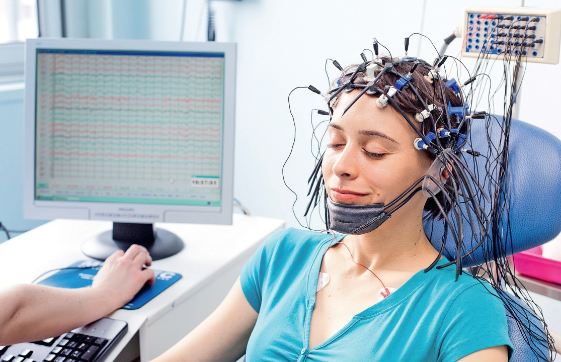
Electroencephalogram (EEG) measures electrical activity to determine neural activation in the brain cortex. It is limited to measure activities in deeper brain regions. Combining EEG with the galvanic skin response (GSR) technique emphasizes the results with the insights provided from deeper and older brain regions. EEG monitors cognitive activities to gather information on how the brain works, which brain regions activated, and how these regions interact without providing insights for behavioral responses. Using EEG with facial expression analysis techniques can determine if subjects express their attitudes and overcome this limitation. It is a non-invasive, non-expensive (compared to other brain imaging tools) and passive recording technique. It directly measured neuronal activities and has a high temporal resolution, making it a good attentional, cognitive, and emotional processing tool for event-related studies. While it has a high temporal resolution, it does not have a good spatial resolution and can not precisely locate the firing neurons (Morin, 2019) (EEG Poket Guide, 2016).
Hsu and Chen set an environment similar to the real world, which is impractical with other brain imaging tools, for their experiment to assess musical stimuli' effects on wine choosing (Hsu & Chen, 2019). However, the EEG tool is lightweight and portable, allowing researchers to collect data in a real-world environment; getting clean data is essential while studying with EEG. Recording data should only reflect brain activity as possible as it can. Electrodes are susceptible, and muscle activities, eye movements, heartbeats, blinks, headset or electrodes movements, line noise, swaying, and swinging can interfere with the EEG technique's results. Because of that, pre-processing and pre-study steps are essential to get qualified results. Electrodes should be placed on clean and dry hair in specific places on the head. Using EEG in multimodal research with other tools like electromyogram, ECG/PPG, and eye-tracking can improve the results and help researchers decrease noises. Denoising the raw signal results requires a certain level of expertise and experience. The interpretation of raw signals can be subjective. Because there is no simple data processing SOP for EEG, it can vary by the respondent population, the recording condition, the stimuli, et cetera. Fortunately, some modern EEG tools can process automatically and generate denoised data. Yadava et al. used a biomedical signal process feature that converts an input signal into a series of small waves by filtering in their study (Morin, 2019) (Yadava, Kumar, Saini, Roy & Prosad, 2017) (EEG Poket Guide, 2016) (Zurawicki, 2010).
Reference
Hsu, L., & Chen, Y. (2019). Music and wine tasting: An experimental neuromarketing study. British Food Journal (1966), 2725-2737.
Imotion EEG Pocket Guide (2016).
Morin, C. (2019). The Ultimate Neuromarketing Research Guide.
Yadava, M., Kumar, P., Saini, R., Roy, P. P., & Prosad Dogra, D. (2017). Analysis of EEG signals and its application to neuromarketing. Multimedia Tools and Applications, 76(18), 19087-19111.
Zurawicki, L. (2010). Neuromarketing. New York: Springer.